SERVICES
RADIATION THERAPY
Radiation therapy, is a type of cancer treatment in which specialists kill cancerous cells in the body by exposing them to ionizing radiation, such as X-rays. It is one of the most widely used cancer treatments, with around half of all patients requiring radiotherapy at some point during the course of the disease.
Cancer is a condition in which cells in a specific part of the body begin to grow and reproduce uncontrollably, forming tumours which affect surrounding tissues and organs and sometimes spread to other parts of the body through the bloodstream or lymphatic system. Radiotherapy involves using carefully selected doses of ionizing radiation to damage the DNA of cancer cells. The DNA controls how they divide. Radiation causes the tumour to shrink and, in some cases, die. It is applied alone or in combination with other treatments, such as chemotherapy or surgery, to cure cancers or control symptoms of the disease.
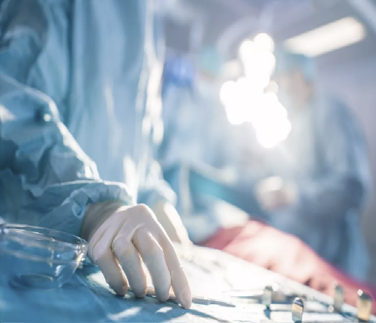
The selected treatment needs a team of qualified experts, consisting of a radiation oncologist, medical physicist and radiation therapy technologist, who use radiation to destroy the tumour while minimizing harm to healthy cells.
Radiotherapy
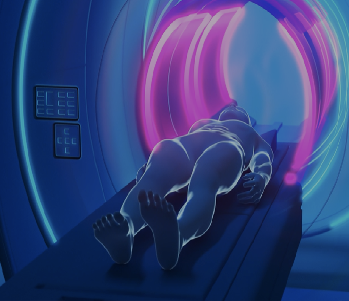
The most common type of radiation therapy is Radiotherapy, whereby radiation is delivered to the tumour area in the form of a high-energy beam. This radiation is emitted from a machine located at a distance from the body, such as a linear accelerator.
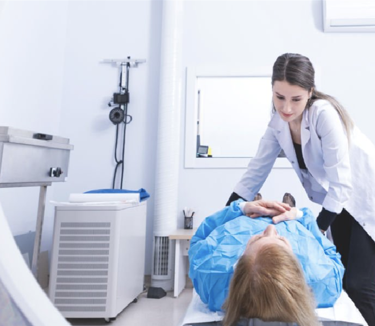
During the therapy, the patient lies motionless on a table while the machine moves around the body, directing precise doses of radiation at the tumour from several angles. The beam size and shape are carefully adjusted to deliver the dose to the tumour while sparing the normal tissues.
Radiotherapy is a highly effective and well-established treatment that millions of patients undergo every year for brain, breast, head and neck, cervical, prostate, skin and other cancers. The effects of the therapy can be seen over time, rather than immediately, and it can take days, weeks or months after the end of the treatment to see the impact of the radiotherapy on the tumour.
Some of the latest advances in radiotherapy, including 3-D conformal radiotherapy, intensity-modulated radiotherapy, VMAT(Volumetric modulated arc therapy technic) and image-guided radiotherapy, help define the target area with high accuracy and effectively deliver precise doses of radiation, causing only minimal damage to healthy cells, tissues and organs. With the VMAT techniq is possible to do the Stereotactic body radiation therapy (SBRT) is an emerging advanced treatment technique that targets tumours with high precision and very high doses of radiation. This delivery method limits the impact on the healthy surrounding tissue, which reduces the likelihood of side effects. As such, it offers a potentially curative therapy or a valuable alternative therapy for many tumour types, including cases in the lungs, liver, brain and pancreas.
In the IBCC center we offer 100% of treatments using the latest technologies such as VMAT and the complex techniques that depend on it such as stereotaxy. We can therefore offer all of our patients high-tech treatments, thus improving the effectiveness of radiotherapy by reducing side effects.
VMAT :
VMAT, or Volumetric Modulated Arc Therapy, is an advanced radiation therapy technique used in the treatment of cancer. It is a form of intensity-modulated radiation therapy (IMRT), which allows for more precise targeting of the cancerous cells while sparing surrounding healthy tissue.
In VMAT, the radiation beam is delivered in an arc around the patient, rather than from fixed angles as in traditional radiotherapy techniques. This arc can vary in shape and size depending on the specific characteristics of the tumor being treated. During treatment, the intensity of the radiation beam is continuously modulated as the machine rotates around the patient, allowing for a highly conformal dose distribution.
The advantages of VMAT over conventional radiotherapy techniques include:
- Increased Treatment Efficiency: VMAT treatments can typically be delivered more quickly than traditional radiotherapy techniques, reducing the overall treatment time for patients.
- Improved Targeting: VMAT allows for better conformality of the radiation dose to the shape of the tumor, minimizing radiation exposure to surrounding healthy tissues and organs.
- Reduced Side Effects: By minimizing radiation exposure to healthy tissues, VMAT can help reduce the risk of side effects and complications associated with radiation therapy.
- Enhanced Patient Comfort: VMAT treatments are typically more comfortable for patients, as they require less time spent immobilized in the treatment position.
- Adaptability: VMAT techniques can be adapted to treat tumors of varying shapes and sizes, making it a versatile option for a wide range of cancer types.
Overall, VMAT represents a significant advancement in the field of radiation therapy, offering improved treatment outcomes and quality of life for cancer patients.
BRACHYTHERAPY
With brachytherapy, a small radioactive source or a XRay source is placed inside the body, enabling a high dose of radiation to be delivered directly to the tumour with only minimal impact on the surrounding tissues.
The source can be placed either. For temporary application, is inserted through a needle or a special applicator. Depending on the dose delivered by the source, the capsule can remain in the body for several minutes to several days. For permanent application, an implant – one example of which is iodine-125 and is usually about the size of a rice grain – is inserted into the body and targets the tumour with radiation, before losing its radioactivity over time.
What are the side effects of radiotherapy?
Side effects depend on the amount of radiation used in radiotherapy and the part of the body that has been irradiated. Patients may experience short- and long-term side effects or no side effects at all.
CHEMOTHERAPY
Chemotherapy is the use of drugs to destroy cancer cells. This type of cancer treatment works by keeping cancer cells from growing, dividing, and making more cells. Chemotherapy is a systemic medication. This means it travels through the bloodstream and reaches all parts of the body.
There are many different kinds of chemotherapy. In general, drugs used for chemotherapy are powerful chemicals that treat cancer by attacking cells during specific parts of the cell cycle. All cells go through the cell cycle, which is how new cells are made. Cancer cells go through this process faster than normal cells, so chemotherapy has more of an effect on these fast-growing cells.

The goals of chemotherapy depend on your type of cancer and how far it has spread. Chemotherapy can be given alone or as a part of a treatment plan that includes different treatments. Some of the ways chemotherapy is used include:
As the primary treatment. Sometimes, the goal of chemotherapy treatment is to get rid of all the cancer and keep it from coming back. This might be called “curative chemotherapy.”
Before other treatments. Chemotherapy can be given before surgery or radiation therapy to shrink tumors. This can be called “neoadjuvant chemotherapy.”
After other treatments. Chemotherapy can be given after surgery or radiation therapy to destroy any remaining cancer cells. This is called “adjuvant chemotherapy.”
To slow the progression of cancer and relieve symptoms. Even when the cancer is not curable, chemotherapy can partially shrink tumors and prevent tumor growth and spread for various lengths of time. In such settings, chemotherapy can extend survival, relieve cancer-related symptoms, and improve quality of life. Chemotherapy used for these purposes is sometimes called “palliative chemotherapy.”
Chemotherapy can be used to treat many types of cancers. It can also be used to treat recurrent cancer and metastatic cancer. Recurrent cancer is cancer that comes back after treatment. Metastatic cancer is cancer that has spread to other parts of the body.
What factors determine a chemotherapy plan?
There are many drugs available to treat cancer. A doctor who specializes in treating cancer with medication is called a medical oncologist. This type of doctor will prescribe your chemotherapy. You may receive a combination of drugs, because this sometimes works better than 1 drug by itself.
The drugs, dose, and treatment schedule depend on many factors. These include:
- The type of cancer
- The stage of the cancer. Cancer stage is determined by the size and location of the tumor and whether or not the cancer has spread. tumor size, its location, and if or where it has spread.
- Your age and general health
- Your body weight
- The possible side effects of each drug. If a drug causes you to have too many side effects, this can also change your treatment plan.
- Any other medical conditions you have
- Previous cancer treatments
Where do you receive chemotherapy?
Chemotherapy can be given at a medical center or taken at home, depending on the specific drug.
Your health care team may need you to come in regularly to the clinic, doctor’s office, or hospital to receive the chemotherapy. This may be called outpatient treatment.
Some types of chemotherapy can be taken at home. Ask your health care team how to safely store, handle, and dispose of your at-home medication. See more below, under “oral chemotherapy” and “topical chemotherapy.”
Learn more about what to expect when getting chemotherapy.
How is chemotherapy delivered?
Chemotherapy may be given in several different ways, which are discussed below.
Intravenous (IV) chemotherapy. Many drugs require injection directly into a vein. This is called intravenous or IV chemotherapy. Treatment takes a few minutes to a few hours. Some IV drugs work better if you get them over a few days or weeks. You take them through a small pump you wear or carry. This is called continuous infusion chemotherapy.
Oral chemotherapy. Oral chemotherapy is taken by mouth. This can be as a pill, capsule, or liquid. This means that you may be able to pick up your medication at the pharmacy and take it at home. Oral treatments for cancer are now more common. Some of these drugs are given daily, and others are given less often. Be sure to ask your health care team about your drug’s schedule and how to store the drug. Learn more about how to keep track of taking your medication at home.
Injected chemotherapy. This is when you receive chemotherapy as a shot. The shot may be given in a muscle or injected under the skin. You may receive these shots in the arm, leg, or abdomen. Abdomen is the medical word for your belly.
Chemotherapy in combination with other cancer treatments
There are other types of drugs besides chemotherapy that also treat cancer, such as hormone therapy, immunotherapy, and targeted therapy. Sometimes oncologists use chemotherapy alongside another type of drug in a person’s treatment plan. These categories of drugs work in different ways to treat cancer, and their side effects are usually different than chemotherapy. Talk with your health care team about what to expect with your specific prescriptions.
Hormone therapy. Hormone therapy is a type of cancer treatment that removes, blocks, or adds specific hormones to the body. It is also called hormonal therapy or endocrine therapy. Hormone therapy can be used to treat several types of cancer.
Immunotherapy. This type of treatment helps your body’s natural defenses fight the cancer. Immunotherapy has developed rapidly during the last few years, and is now an important part of treatment for several types of cancer.
Targeted therapy. These treatments target and disable genes or proteins found in cancer cells that the cancer cells need to grow. Targeted therapy can treat many types of cancer.
How long will I need chemotherapy?
Chemotherapy is often given for a specific time, such as 6 months or a year. Or you might receive chemotherapy for as long as it works.
Side effects from many anti-cancer drugs are too severe to give treatment every day. Doctors usually give these drugs with breaks, so you have time to rest and recover before the next treatment. This lets your healthy cells heal.
For example, you might get a dose of chemotherapy on the first day and then have 3 weeks of recovery time before repeating the treatment. Each 3-week period is called a treatment cycle. Several cycles make up a course of chemotherapy. A course usually lasts 3 months or more.
Some cancers are treated with less recovery time between cycles. This is called a dose-dense schedule. It can make chemotherapy more effective against some cancers. But it also increases the risk of side effects.
Your health care team will explain how often and for how long you’ll receive chemotherapy. Be sure to talk with your doctor, nurse, or other team member regularly about side effects of chemotherapy, including what you can expect and what you are experiencing.
MEDICAL IMAGING
Evaluation of disease location and spread
Determining whether the cancer is at an early stage or has spread to other parts of the body is done through medical imaging. Image-guided procedures such as biopsies are minimally invasive and are necessary for accurate tissue diagnosis.
Medical imaging modalities, such as computed tomography (CT), ultrasound, magnetic resonance imaging (MRI) and positron emission tomography (PET), are essential for accurate cancer diagnosis and staging. These techniques help identify the location, size and extent of the tumour, as well as its relationship to adjacent structures and the presence of metastases.
Diagnostic imaging describes various techniques of viewing the inside of the body to help figure out the causes of an illness or injury and confirm a diagnosis. Doctors also use it to see how well a patient’s body responds to treatment for a fracture or illness.
Medical imaging plays a crucial role in cancer patient management and is requisite for the planning, delivery and evaluation of radiotherapy treatment. The integration of advanced imaging techniques in radiotherapy has revolutionised cancer care and improved patient outcomes. In this context, the role of medical imaging includes:
Physicians also refer to a CT scan as a “cat scan.” The test combines a string of X-ray scans or images taken from various angles. Computer software then generates cross-sectional images (slices) of blood vessels and soft tissues inside the body. CT scans can offer a more thorough picture than standard X-rays. They’re frequently used to quickly examine individuals who have internal injuries from a trauma.
Doctors can use CT scans to evaluate the spine, brain, abdomen, neck and chest. They provide clear images of both hard and soft tissues. The pictures the CT scans produce allow doctors to quickly make medical decisions if required. Because of this quality, CT scans are commonly performed in both imaging centers and hospitals. They help physicians find injuries and diseases that could previously only be found in a surgery or autopsy. While CT scans use low doses of radiation, they’re still relatively non-invasive and safe.
These scans are useful in a variety of medical situations where diagnostic imagery is required. They can assess slight abnormalities in soft tissue like the brain as well as other organs. Doctors also use the images when patients have certain symptoms like dizziness or pain. They’re even useful in examining the spread of certain conditions, such as cancer. Depending on where the technologist directs the CT scan in your body, there are various uses for it, including:
- Brain or head CT scans:Check for stroke, bleeds, masses and other abnormalities and examine the skull
- Chest CT scans:Provide further insight into abnormalities after a standard chest x-ray
- Neck CT scans:Look for enlarged glands or lymph nodes and study lumps.
- Spine CT scans:Detect spine problems like spinal canal narrowing, a herniated disc or fractures
- Sinus CT scans:Detect and diagnose obstructions or sinus disease
- Pelvic or abnormal CT scans:Check organs in this area and diagnose unexplained pain in the abdomen
Treatment planning for Radiotherapy
Imaging is used for treatment planning: from medication and radiotherapy to surgery or, in certain cases, palliation. During radiotherapy planning, medical imaging generates three-dimensional images of the tumour, allowing for better radiation therapy targeting of the tumour while minimising damage to healthy tissues.
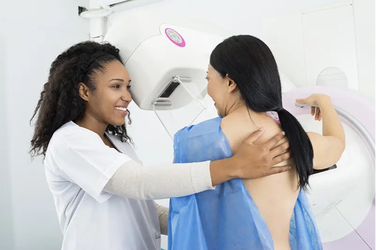
Mammograms
Mammograms are a type of x-ray image of the breasts. They check for early breast cancer signs like small lumps you or your doctor can’t feel through the use of a low-dose x-ray. Mammograms also show breast tissue changes that could be a sign of early-stage breast cancer.
A radiologist uses digital mammography to identify and diagnose cancer nodules that older systems can’t detect. Mammograms are the best way to detect early breast cancer because — in some cases as much as a few years before you can feel it. Having mammograms regularly comes with various benefits, including the following:
- They detect breast cancer early on, which saves lives.
- They reduce your risk of dying because of breast cancer by 30 percent.
- Getting treatment early means you can keep your breasts and don’t have to resort to mastectomy.
Mammograms, to some women, can be uncomfortable and sometimes even painful. A lot of this feeling depends on how big your breasts are and the amount of pressure needed to be applied. The discomfort will last only a few minutes, and the fact that this procedure can save your life makes it worth it.
Ultrasound
Also referred to as “sonography,” ultrasound imaging is a safe imaging method that creates images of the inside of the body. It doesn’t use radiation, but rather high-frequency waves. As a result, it’s a safe procedure during pregnancy. The ultrasound images are in real-time and show the structure and movement of internal organs and the blood flow through vessels.
During an ultrasound, a sonographer will hold a transducer — a handheld device — over your skin. Sometimes it’s placed internally. It uses sound waves traveling through soft tissue and fluids, and as it hits denser surfaces, it echoes or bounces back, which is how the images are created. More ultrasound echoes back when the object is denser.
Doctors can diagnose a large variety of health conditions with an ultrasound. The images it creates also help physicians come up with treatment plans. If you have symptoms such as swelling, infection or pain, your doctor might suggest an ultrasound to determine the cause. Ultrasounds are also used to assist anesthesiologist during surgical procedures when they’re guiding needles near nerves.
In a lot of cases, ultrasounds are tools that allow doctors to examine problems related to abdominal issues, circulation, urology, obstetrics, newborn care and even musculoskeletal conditions. Some common body parts physicians use ultrasounds for include:
- Heart
- Joints
- Uterus
- Blood vessels
- Muscles
- Bladder
- Kidneys
- And more
X-ray
X-rays are among the most commonly used and well-known diagnostic imaging tests. Doctors use them to view the inside of the body. X-ray equipment generates a high-energy beam that dense tissue and bones can’t absorb, but that passes through other areas of the body. This process generates an image, allowing your doctor to see if you suffered an injury to your bones.
SERVICES
Radiation therapy, is a type of cancer treatment in which specialists kill cancerous cells in the body by exposing them to ionizing radiation, such as X-rays. It is one of the most widely used cancer treatments, with around half of all patients requiring radiotherapy at some point during the course of the disease.
Cancer is a condition in which cells in a specific part of the body begin to grow and reproduce uncontrollably, forming tumours which affect surrounding tissues and organs and sometimes spread to other parts of the body through the bloodstream or lymphatic system. Radiotherapy involves using carefully selected doses of ionizing radiation to damage the DNA of cancer cells. The DNA controls how they divide. Radiation causes the tumour to shrink and, in some cases, die. It is applied alone or in combination with other treatments, such as chemotherapy or surgery, to cure cancers or control symptoms of the disease.
RADIATION THERAPY
The selected treatment needs a team of qualified experts, consisting of a radiation oncologist, medical physicist and radiation therapy technologist, who use radiation to destroy the tumour while minimizing harm to healthy cells.
Radiotherapy
The most common type of radiation therapy is Radiotherapy, whereby radiation is delivered to the tumour area in the form of a high-energy beam. This radiation is emitted from a machine located at a distance from the body, such as a linear accelerator.
During the therapy, the patient lies motionless on a table while the machine moves around the body, directing precise doses of radiation at the tumour from several angles. The beam size and shape are carefully adjusted to deliver the dose to the tumour while sparing the normal tissues.
Radiotherapy is a highly effective and well-established treatment that millions of patients undergo every year for brain, breast, head and neck, cervical, prostate, skin and other cancers. The effects of the therapy can be seen over time, rather than immediately, and it can take days, weeks or months after the end of the treatment to see the impact of the radiotherapy on the tumour.
Some of the latest advances in radiotherapy, including 3-D conformal radiotherapy, intensity-modulated radiotherapy, VMAT(Volumetric modulated arc therapy technic) and image-guided radiotherapy, help define the target area with high accuracy and effectively deliver precise doses of radiation, causing only minimal damage to healthy cells, tissues and organs. With the VMAT techniq is possible to do the Stereotactic body radiation therapy (SBRT) is an emerging advanced treatment technique that targets tumours with high precision and very high doses of radiation. This delivery method limits the impact on the healthy surrounding tissue, which reduces the likelihood of side effects. As such, it offers a potentially curative therapy or a valuable alternative therapy for many tumour types, including cases in the lungs, liver, brain and pancreas.
In the IBCC center we offer 100% of treatments using the latest technologies such as VMAT and the complex techniques that depend on it such as stereotaxy. We can therefore offer all of our patients high-tech treatments, thus improving the effectiveness of radiotherapy by reducing side effects.
VMAT :
VMAT, or Volumetric Modulated Arc Therapy, is an advanced radiation therapy technique used in the treatment of cancer. It is a form of intensity-modulated radiation therapy (IMRT), which allows for more precise targeting of the cancerous cells while sparing surrounding healthy tissue.
In VMAT, the radiation beam is delivered in an arc around the patient, rather than from fixed angles as in traditional radiotherapy techniques. This arc can vary in shape and size depending on the specific characteristics of the tumor being treated. During treatment, the intensity of the radiation beam is continuously modulated as the machine rotates around the patient, allowing for a highly conformal dose distribution.
The advantages of VMAT over conventional radiotherapy techniques include:
- Increased Treatment Efficiency: VMAT treatments can typically be delivered more quickly than traditional radiotherapy techniques, reducing the overall treatment time for patients.
- Improved Targeting: VMAT allows for better conformality of the radiation dose to the shape of the tumor, minimizing radiation exposure to surrounding healthy tissues and organs.
- Reduced Side Effects: By minimizing radiation exposure to healthy tissues, VMAT can help reduce the risk of side effects and complications associated with radiation therapy.
- Enhanced Patient Comfort: VMAT treatments are typically more comfortable for patients, as they require less time spent immobilized in the treatment position.
- Adaptability: VMAT techniques can be adapted to treat tumors of varying shapes and sizes, making it a versatile option for a wide range of cancer types.
Overall, VMAT represents a significant advancement in the field of radiation therapy, offering improved treatment outcomes and quality of life for cancer patients.
BRACHYTHERAPY
With brachytherapy, a small radioactive source or a XRay source is placed inside the body, enabling a high dose of radiation to be delivered directly to the tumour with only minimal impact on the surrounding tissues.
The source can be placed either. For temporary application, is inserted through a needle or a special applicator. Depending on the dose delivered by the source, the capsule can remain in the body for several minutes to several days. For permanent application, an implant – one example of which is iodine-125 and is usually about the size of a rice grain – is inserted into the body and targets the tumour with radiation, before losing its radioactivity over time.
What are the side effects of radiotherapy?
Side effects depend on the amount of radiation used in radiotherapy and the part of the body that has been irradiated. Patients may experience short- and long-term side effects or no side effects at all.
CHEMOTHERAPY
Chemotherapy is the use of drugs to destroy cancer cells. This type of cancer treatment works by keeping cancer cells from growing, dividing, and making more cells. Chemotherapy is a systemic medication. This means it travels through the bloodstream and reaches all parts of the body.
There are many different kinds of chemotherapy. In general, drugs used for chemotherapy are powerful chemicals that treat cancer by attacking cells during specific parts of the cell cycle. All cells go through the cell cycle, which is how new cells are made. Cancer cells go through this process faster than normal cells, so chemotherapy has more of an effect on these fast-growing cells.
The goals of chemotherapy depend on your type of cancer and how far it has spread. Chemotherapy can be given alone or as a part of a treatment plan that includes different treatments. Some of the ways chemotherapy is used include:
As the primary treatment. Sometimes, the goal of chemotherapy treatment is to get rid of all the cancer and keep it from coming back. This might be called “curative chemotherapy.”
Before other treatments. Chemotherapy can be given before surgery or radiation therapy to shrink tumors. This can be called “neoadjuvant chemotherapy.”
After other treatments. Chemotherapy can be given after surgery or radiation therapy to destroy any remaining cancer cells. This is called “adjuvant chemotherapy.”
To slow the progression of cancer and relieve symptoms. Even when the cancer is not curable, chemotherapy can partially shrink tumors and prevent tumor growth and spread for various lengths of time. In such settings, chemotherapy can extend survival, relieve cancer-related symptoms, and improve quality of life. Chemotherapy used for these purposes is sometimes called “palliative chemotherapy.”
Chemotherapy can be used to treat many types of cancers. It can also be used to treat recurrent cancer and metastatic cancer. Recurrent cancer is cancer that comes back after treatment. Metastatic cancer is cancer that has spread to other parts of the body.
What factors determine a chemotherapy plan?
There are many drugs available to treat cancer. A doctor who specializes in treating cancer with medication is called a medical oncologist. This type of doctor will prescribe your chemotherapy. You may receive a combination of drugs, because this sometimes works better than 1 drug by itself.
The drugs, dose, and treatment schedule depend on many factors. These include:
- The type of cancer
- The stage of the cancer. Cancer stage is determined by the size and location of the tumor and whether or not the cancer has spread. tumor size, its location, and if or where it has spread.
- Your age and general health
- Your body weight
- The possible side effects of each drug. If a drug causes you to have too many side effects, this can also change your treatment plan.
- Any other medical conditions you have
- Previous cancer treatments
Where do you receive chemotherapy?
Chemotherapy can be given at a medical center or taken at home, depending on the specific drug.
Your health care team may need you to come in regularly to the clinic, doctor’s office, or hospital to receive the chemotherapy. This may be called outpatient treatment.
Some types of chemotherapy can be taken at home. Ask your health care team how to safely store, handle, and dispose of your at-home medication. See more below, under “oral chemotherapy” and “topical chemotherapy.”
Learn more about what to expect when getting chemotherapy.
How is chemotherapy delivered?
Chemotherapy may be given in several different ways, which are discussed below.
Intravenous (IV) chemotherapy. Many drugs require injection directly into a vein. This is called intravenous or IV chemotherapy. Treatment takes a few minutes to a few hours. Some IV drugs work better if you get them over a few days or weeks. You take them through a small pump you wear or carry. This is called continuous infusion chemotherapy.
Oral chemotherapy. Oral chemotherapy is taken by mouth. This can be as a pill, capsule, or liquid. This means that you may be able to pick up your medication at the pharmacy and take it at home. Oral treatments for cancer are now more common. Some of these drugs are given daily, and others are given less often. Be sure to ask your health care team about your drug’s schedule and how to store the drug. Learn more about how to keep track of taking your medication at home.
Injected chemotherapy. This is when you receive chemotherapy as a shot. The shot may be given in a muscle or injected under the skin. You may receive these shots in the arm, leg, or abdomen. Abdomen is the medical word for your belly.
Chemotherapy in combination with other cancer treatments
There are other types of drugs besides chemotherapy that also treat cancer, such as hormone therapy, immunotherapy, and targeted therapy. Sometimes oncologists use chemotherapy alongside another type of drug in a person’s treatment plan. These categories of drugs work in different ways to treat cancer, and their side effects are usually different than chemotherapy. Talk with your health care team about what to expect with your specific prescriptions.
Hormone therapy. Hormone therapy is a type of cancer treatment that removes, blocks, or adds specific hormones to the body. It is also called hormonal therapy or endocrine therapy. Hormone therapy can be used to treat several types of cancer.
Immunotherapy. This type of treatment helps your body’s natural defenses fight the cancer. Immunotherapy has developed rapidly during the last few years, and is now an important part of treatment for several types of cancer.
Targeted therapy. These treatments target and disable genes or proteins found in cancer cells that the cancer cells need to grow. Targeted therapy can treat many types of cancer.
How long will I need chemotherapy?
Chemotherapy is often given for a specific time, such as 6 months or a year. Or you might receive chemotherapy for as long as it works.
Side effects from many anti-cancer drugs are too severe to give treatment every day. Doctors usually give these drugs with breaks, so you have time to rest and recover before the next treatment. This lets your healthy cells heal.
For example, you might get a dose of chemotherapy on the first day and then have 3 weeks of recovery time before repeating the treatment. Each 3-week period is called a treatment cycle. Several cycles make up a course of chemotherapy. A course usually lasts 3 months or more.
Some cancers are treated with less recovery time between cycles. This is called a dose-dense schedule. It can make chemotherapy more effective against some cancers. But it also increases the risk of side effects.
Your health care team will explain how often and for how long you’ll receive chemotherapy. Be sure to talk with your doctor, nurse, or other team member regularly about side effects of chemotherapy, including what you can expect and what you are experiencing.
MEDICAL IMAGING
Evaluation of disease location and spread
Determining whether the cancer is at an early stage or has spread to other parts of the body is done through medical imaging. Image-guided procedures such as biopsies are minimally invasive and are necessary for accurate tissue diagnosis.
Medical imaging modalities, such as computed tomography (CT), ultrasound, magnetic resonance imaging (MRI) and positron emission tomography (PET), are essential for accurate cancer diagnosis and staging. These techniques help identify the location, size and extent of the tumour, as well as its relationship to adjacent structures and the presence of metastases.
Diagnostic imaging describes various techniques of viewing the inside of the body to help figure out the causes of an illness or injury and confirm a diagnosis. Doctors also use it to see how well a patient’s body responds to treatment for a fracture or illness.
Medical imaging plays a crucial role in cancer patient management and is requisite for the planning, delivery and evaluation of radiotherapy treatment. The integration of advanced imaging techniques in radiotherapy has revolutionised cancer care and improved patient outcomes. In this context, the role of medical imaging includes:
Physicians also refer to a CT scan as a “cat scan.” The test combines a string of X-ray scans or images taken from various angles. Computer software then generates cross-sectional images (slices) of blood vessels and soft tissues inside the body. CT scans can offer a more thorough picture than standard X-rays. They’re frequently used to quickly examine individuals who have internal injuries from a trauma.
Doctors can use CT scans to evaluate the spine, brain, abdomen, neck and chest. They provide clear images of both hard and soft tissues. The pictures the CT scans produce allow doctors to quickly make medical decisions if required. Because of this quality, CT scans are commonly performed in both imaging centers and hospitals. They help physicians find injuries and diseases that could previously only be found in a surgery or autopsy. While CT scans use low doses of radiation, they’re still relatively non-invasive and safe.
These scans are useful in a variety of medical situations where diagnostic imagery is required. They can assess slight abnormalities in soft tissue like the brain as well as other organs. Doctors also use the images when patients have certain symptoms like dizziness or pain. They’re even useful in examining the spread of certain conditions, such as cancer. Depending on where the technologist directs the CT scan in your body, there are various uses for it, including:
- Brain or head CT scans:Check for stroke, bleeds, masses and other abnormalities and examine the skull
- Chest CT scans:Provide further insight into abnormalities after a standard chest x-ray
- Neck CT scans:Look for enlarged glands or lymph nodes and study lumps.
- Spine CT scans:Detect spine problems like spinal canal narrowing, a herniated disc or fractures
- Sinus CT scans:Detect and diagnose obstructions or sinus disease
- Pelvic or abnormal CT scans:Check organs in this area and diagnose unexplained pain in the abdomen
Treatment planning for Radiotherapy
Imaging is used for treatment planning: from medication and radiotherapy to surgery or, in certain cases, palliation. During radiotherapy planning, medical imaging generates three-dimensional images of the tumour, allowing for better radiation therapy targeting of the tumour while minimising damage to healthy tissues.
Mammograms
Mammograms are a type of x-ray image of the breasts. They check for early breast cancer signs like small lumps you or your doctor can’t feel through the use of a low-dose x-ray. Mammograms also show breast tissue changes that could be a sign of early-stage breast cancer.
A radiologist uses digital mammography to identify and diagnose cancer nodules that older systems can’t detect. Mammograms are the best way to detect early breast cancer because — in some cases as much as a few years before you can feel it. Having mammograms regularly comes with various benefits, including the following:
- They detect breast cancer early on, which saves lives.
- They reduce your risk of dying because of breast cancer by 30 percent.
- Getting treatment early means you can keep your breasts and don’t have to resort to mastectomy.
Mammograms, to some women, can be uncomfortable and sometimes even painful. A lot of this feeling depends on how big your breasts are and the amount of pressure needed to be applied. The discomfort will last only a few minutes, and the fact that this procedure can save your life makes it worth it.
Ultrasound
Also referred to as “sonography,” ultrasound imaging is a safe imaging method that creates images of the inside of the body. It doesn’t use radiation, but rather high-frequency waves. As a result, it’s a safe procedure during pregnancy. The ultrasound images are in real-time and show the structure and movement of internal organs and the blood flow through vessels.
During an ultrasound, a sonographer will hold a transducer — a handheld device — over your skin. Sometimes it’s placed internally. It uses sound waves traveling through soft tissue and fluids, and as it hits denser surfaces, it echoes or bounces back, which is how the images are created. More ultrasound echoes back when the object is denser.
Doctors can diagnose a large variety of health conditions with an ultrasound. The images it creates also help physicians come up with treatment plans. If you have symptoms such as swelling, infection or pain, your doctor might suggest an ultrasound to determine the cause. Ultrasounds are also used to assist anesthesiologist during surgical procedures when they’re guiding needles near nerves.
In a lot of cases, ultrasounds are tools that allow doctors to examine problems related to abdominal issues, circulation, urology, obstetrics, newborn care and even musculoskeletal conditions. Some common body parts physicians use ultrasounds for include:
- Heart
- Joints
- Uterus
- Blood vessels
- Muscles
- Bladder
- Kidneys
- And more
X-ray
X-rays are among the most commonly used and well-known diagnostic imaging tests. Doctors use them to view the inside of the body. X-ray equipment generates a high-energy beam that dense tissue and bones can’t absorb, but that passes through other areas of the body. This process generates an image, allowing your doctor to see if you suffered an injury to your bones.